|
 |
 |
|
|
|
|
Dr. William Bowers
Edited and
illustrated by Dr Richard Hunt
Medical Microbiology
(MBIM 650/720)
SUGGESTED READING:
Roitt, Brostoff, Male, 6th Edition, Mosby, 2001 Chapter 8, pp. 132-136; Chapter
7, pp. 119-129 |
IMMUNOLOGY - CHAPTER TWELVE
CELL-MEDIATED IMMUNITY:
Cell interactions in specific immune responses
|
|
TEACHING
OBJECTIVES
Helper
T cell-B cell interactions for antibody formation against hapten-conjugated
proteins and complex proteins
Thymus-independent
antigens
Properties
and functions of cytokines
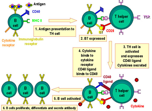
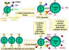 Figure 1
Molecules involved in the interactions of B and TH cells
Antigen  is processed by B cell.
Co-stimulators are expressed. The processed antigen peptide is processed by B cell.
Co-stimulators are expressed. The processed antigen peptide
 is presented in association with MHC class II antigens. The T cell
recognizes the peptide along with the MHC antigen and the
co-stimulators. The T cell expresses CD40 ligand. The latter binds
to CD40 antigen on the B cell and the B cells divide and
differentiate. Antibodies are produced by the B cell
is presented in association with MHC class II antigens. The T cell
recognizes the peptide along with the MHC antigen and the
co-stimulators. The T cell expresses CD40 ligand. The latter binds
to CD40 antigen on the B cell and the B cells divide and
differentiate. Antibodies are produced by the B cell |
I. HELPER T CELL-B CELL
INTERACTIONS IN ANTIBODY FORMATION
A. Hapten-carrier effect
Historically one of the
major findings was that T cells and B cells are required in order to produce
antibody to a complex protein. A major contribution to our understanding of
this process came from studies on the formation of anti-hapten antibodies.
Recall that a hapten
injected by itself cannot elicit an antibody response. Rather antibodies
against haptens require that the hapten be conjugated to a protein (sometimes
termed a carrier).
These studies with hapten-carrier established that:
1. Th cells recognize
the carrier, and B cells recognize hapten.
2. There must be
cooperation between hapten-specific B cells and protein (carrier)-specific
helper T cells.
3. Interaction between
the hapten-specific B cell and the carrier-specific helper T cell are class
II self MHC-restricted. The helper T cell cooperates only with B cells that
express class II MHC molecules recognized as self by the T cells.
B. B cells as antigen
presenting cells
B cells occupy a
unique position in immune responses because they express immunoglobulin (Ig)
and class II MHC molecules on their cell surface. They therefore are capable
of producing antibody having the same specificity as that expressed by their
immunoglobulin receptor; in addition they can function as an antigen
presenting cell. In terms of the hapten-carrier protein findings, the
mechanism is thought to be the following: the hapten is recognized by the Ig
receptor,
the hapten-carrier brought into the B cell, processed, and peptide fragments
of the carrier protein presented to a helper T cell. Activation of the T
cell results in the production of cytokines that enable the hapten-specific
B cell to become activated to produce soluble anti-hapten antibodies. Figure
1 summarizes the B cell-T cell interactions that occur.
Note that there are
multiple signals delivered to the B cells in this model of Th cell-B cell
interaction. As was the case for activation of T cells where the signal
derived from the TCR recognition of a peptide-MHC molecule was by itself
insufficient for T cell activation, so too for the B cell. Binding of an
antigen to the immunoglobulin receptor delivers one signal to the B cell,
but that is insufficient. Second signals delivered by costimulatory
molecules are required; the most important of these is CD40L on the T cell
that binds to CD40 on the B cell to initiate delivery of a second signal.
|
 Figure 2
Cooperation of cells in the immune response
Antigen-presenting cells (e.g. dendritic cells) present processed
antigen to virgin T cells, thereby priming them. B cells also process
the antigen and present it to the T cells. They then receive signals
from the T cells that cause them to divide and differentiate. Some B
cells form antibody-forming cells while a few form B memory cells |
C. Extension of this
model to complex protein antigens (T-dependent antigens)
The same mechanism
described above can cover all multideterminant complex protein antigens that
require helper T cells. These antigens are referred to as thymus-dependent
antigens. If one determinant is recognized by B cells (analogous to the hapten)
and the same or different determinant is recognized by the helper T cells
(analogous to the carrier), the same model applies. This is shown in Figure 2.
D. B cells in secondary
responses
As a consequence of a
primary response, many memory B cells are created. These carry a high-affinity
receptor, Ig, which allows them to bind and present antigen at much lower
concentrations than is required for macrophages or dendritic cells.
II. THYMUS-INDEPENDENT
ANTIGENS
The thymus-independent
antigens (T-independent antigens) are those that produce normal antibody
responses in athymic (thymus-less or nude) mice, i.e. under conditions where T
cells are absent. T-independent antigens have the following properties:
1. activate B cells at
high concentrations, i.e. are polyclonal B cell activators (antigens like
lipopolysaccharide, LPS, sometimes termed B cell mitogens).
2. are large polymeric
molecules with repeating antigenic determinants.
3. are particularly
resistant to degradation
4. Some antigens
activate both immature and mature B cells; other antigens activate only
mature B cells and are thus not especially effective in infants where B
cells are mostly immature.
5.
Responses to several
T-independent antigens are
dominated by CD5 B cells,
described below.
Unlike the
thymus-dependent antigens, the thymus-independent antigens:
1. do not produce
isotype switching (IgM is almost exclusively produced)
2. do not demonstrate
affinity maturation (in which antibodies of progressively higher affinity
are produced)
3. do not show
secondary responses (no memory B cells).
The thymus-independent
antigen pathway is important because humoral immunity is the major mechanism
of defense against many harmful bacteria that have polysaccharides in their
cell wall. Individuals with depressed T cell systems can still resist these
types of bacterial infections.
|
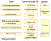 Figure 4
Origin of B cell tumors. These tumors arise as clonal outgrowths from
normal B cells at different developmental stages. The tumor cells behave
in a similar manner to their normal equivalent and go to similar parts
of the body |
III. CD5+ B
CELLS
CD5+ B cells
(sometimes referred to as B-1 cells) form a population that is distinct from
conventional B cells (sometimes referred to as B-2 cells). They have the
following characteristics:
1. are the first B
cells to appear in ontogeny
2. express surface IgM,
but little or no IgD
3. produce immunoglobulins, mainly IgM, from unmutated or minimally mutated germline
genes
4. produce antibodies
of low avidity that are polyreactive (i.e., bind multiple different
antigens, mainly bacterial polysaccharides and double stranded DNA)
5. contribute most of
the IgM found in adult serum
6. do not develop into
memory cells
7. are self-renewing
in adults (i.e., do not continue to arise from a stem cell in the bone
marrow as do conventional B cells)
8. reside in
peripheral tissues and are the predominant lymphocyte in the peritoneal
cavity.
The following table
contrasts CD5+ B cells with conventional B cells.
|
Table 1
- COMPARISON OF PROPERTIES OF
CD5+ B CELLS AND CONVENTIONAL B CELLS
|
|
Properties |
CD5+ B
cells |
Conventional B Cells
|
|
Ontogeny |
Early |
Late |
|
Renewal |
Self Renewal |
Replaced from bone
marrow |
|
Production of
Immunoglobulin |
High |
Low |
|
Isotypes secreted |
IgM>>IgG |
IgG>IgM |
|
Bind multiple
different ligands |
Yes |
No |
|
Adapted from Janeway
and Travers, Immunobiology
|
As shown in Figure 4, tumors
can arise from CD5+ B cells and conventional B cells at various
stages in their development.
|
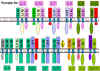 Figure 5 Receptors for various cytokines showing common subunits
 |
IV. CYTOKINES
Cytokines
are a diverse group of non-antibody proteins released by cells that act as
intercellular mediators, especially in immune processes.
A. Cytokines are clinically
important as biological response modifiers. Terms in the literature:
1. Monokines - produced by
mononuclear phagocytes
2. Lymphokines -
produced by activated T cells, primarily helper T cells
3. Interleukins - name
given to many cytokines, abbreviated as IL and given a number
B. Properties
1. Produced by cells involved
in both natural and specific immunity
2. Mediate and regulate immune and
inflammatory responses
3. Secretion is brief and limited
a. Cytokines are not stored as pre-formed
molecules
b. Synthesis is initiated by new short-lived
gene transcription
c. mRNA is short-lived
d. This results in production of cytokine as
needed
4. Many individual cytokines are produced
by many cell types and act on many cell types (they are pleiotropic)
5. In many cases cytokines have similar
actions (they are redundant). Redundancy is due to the following: Receptors
for cytokines are heterodimers (sometimes heterotrimers) that can be grouped
into families in which one subunit is common to all members of a given family.
Some examples are shown in Figure 5.
Since the subunit common to all members of the family
functions in binding cytokine and in signal transduction, a receptor for one
cytokine can often respond to another cytokine in the same family. Thus, an
individual lacking IL-2, for example, is not adversely affected because other
cytokines (IL-15, IL-7, IL-9, etc.) assume its function. Similarly, a mutation
in a cytokine receptor subunit other than the one in common often has little
effect. On the other hand, a mutation in the common subunit has profound
effects. Again, as an example, mutation in the gene for the IL-2Rgamma
subunit causes human X-linked severe combined immunodeficiency (XSCID)
characterized by a complete or nearly complete T cell defect.
6. Often influence the synthesis of other
cytokines
a. They can produce cascades, or enhance or
suppress production of other cytokines
b. They exert positive or negative regulatory
mechanisms for immune and inflammatory responses
7. Often influence the action of other
cytokines. Effects can be:
a. antagonistic
b. additive
c. greater than additive (synergistic)
8. Bind to specific receptors on
target cells with high affinity. Compare with antigen binding to antibody
or peptide binding to a MHC molecule which both show much lower binding
affinities.
9. Cells that can respond to a cytokine
are:
a. same cell that secreted cytokine:
autocrine
b. a nearby cell: paracrine
c. a distant cell reached through the
circulation: endocrine
10. Cellular responses to cytokines are
generally slow (hours), require new mRNA and protein synthesis
C. Cytokines can be grouped according to
function
1. Mediators and regulators of Natural
Immunity
Tumor
Necrosis Factor (TNF)
Interleukin-1 (IL-1)
Chemokines
Interleukin-10
Interferon-gamma (IFN-gamma)
2. Mediators and regulators of specific
immunity
Interleukin-2
(IL-2)
Interleukin-4 (IL-4)
Interleukin-5 (IL-5)
Interleukin-10 (IL-10)
Interferon-gamma (IFN-gamma)
3. Stimulators of hematopoeisis
Interleukin-3
(IL-3)
Colony-Stimulating Factors (CSFs)
|
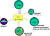 Figure 6
Immuno-regulatory actions of interleukin-2
 Figure 7
T cell proliferation and cytokines. When T cells are resting, they
do not make cytokines such as interleukins 2, 4 or 7. Nor do they
express large amounts of their receptors. There are no IL-2
receptors. Activation of T cells results in the formation of high
affinity IL-2 receptors and induction of the synthesis and
secretion of IL-2 and Il-4. These bind to their receptors and the
cells proliferate. When stimulation by interleukins declines (e.g.
when antigen stimulation declines), receptors decay and the
proliferative phase is at an end. Note: stimulation by the
cytokines can be paracrine
or autocrine
|
D. Functions of selected cytokines:
Mediators and regulators of natural immunity
1.
Tumor Necrosis Factor (TNF) also called TNF-gamma
a)
is produced by activated macrophages
b) is the most important
mediator of acute inflammation in response to Gram-negative bacteria and
other infectious microbes
c) mediates the recruitment
of polymorphonuclear leukocytes (PMNs) and monocytes to the
site of infection:
i)
stimulates endothelial cells to express new adhesion molecules that
make the cell surface "sticky" for PMN and monocytes
ii)
stimulates endothelial cells and macrophages to produce chemokines that
induce leukocyte chemotaxis and
recruitment
d) acts on the hypothalamus to produce fever
e) promotes the
production of acute phase proteins by the liver
2. Interleukin-1
a)
produced by activated macrophages
b) effects are similar to
those of TNF
3.
Chemokines
The name chemokine is a contraction
of chemotactic cytokines
a)
These are are a large family of substances (more than 50) produced by many different
leukocytes and tissue cells
b) They recruit
leukocytes to sites of infection
c) They play a
role in lymphocyte trafficking
4.
Interleukin-10
a)
is produced by activated macrophages
b)
acts as an inhibitor of activated macrophages by blocking production of
TNF
E.
Functions of Selected Cytokines: Mediators and Regulators of
Specific Immunity
1.
Interleukin-2
a)
is produced mainly by helper T cells (CD4+); less by cytoxic
T cells (CD8+)
b) mainly
functions to promote T cell division and to increase production of
other cytokines
c) has other
functions that are shown in Figure 6
d) has autocrine functions on T cell proliferation as depicted in
Figure 7
2. Interleukin-4
a)
is produced mainly by Th2 subpopulation of helper T cells
(CD4+). RECALL that Th2
cells are required for antibody production by B cells
b) stimulates
immunoglobulin class switching to the IgE isotype.
(IgE is involved in eosinophil-mediated elimination of
helminths and arthropods.)
c)
stimulates development of Th2 cells from naive CD4+ T cells
d)
promotes growth of differentiated Th2 cells
3.
Interleukin-5
a)
is produced mainly by the Th2 subpopulation of helper T cells
(CD4+)
b) promotes
growth and differentiation of eosinophils
c) activates
mature eosinophils
IL-4
and IL-5 function together
IgE opsonizes helminths that then bind to eosinophils which upon
activation kill the helminth.
|
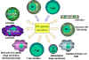 Figure 8
Immunoregulatory actions of interferon gamma on the immune
system. Note the anti-proliferation and anti-viral activities are
weaker than those of IFN alpha and IFN beta. IFN gamma is the most
potent of the three at macrophage activation and in inducing class
II MHC expression |
4.
Interferons (IFN)
There are three
groups of interferons: IFN-alpha , IFN-beta , IFN-gamma
a)
IFN-alpha: Twenty
variants are produced by leukocytes in response to viruses
b) IFN-beta: This
is a single protein produced by fibroblasts and other cells in
response to viruses
Both
IFN-alpha and IFN-beta
inhibit viral replication and increase expression of class I
MHC on cells
c)
IFN-gamma:
i) This protein is produced
by the Th1 subpopulation of helper T cells (CD4+), cytotoxic T
cells (CD8+), and NK cells.
RECALL that Th1 cells are involved in the elimination of pathogens
residing intracellularly in vesicular compartments.
ii)
IFN-gamma functions in both natural and specific immunity
Natural
Immunity
- IFN-gamma enhances
the microbicidal function of macrophages through formation of
nitric oxide and reactive oxygen intermediates (ROI)
Specific
Immunity
- IFN-gamma stimulates
the expression of class I and class II MHC molecules and
co-stimulatory molecules on antigen presenting cells
- IFN-gamma promotes the
differentiation of naive helper T cells into Th1 cells
- IFN-gamma activates
polymorphonuclear leukocytes (PMN) and cytotoxic T cells and
increases the cytotoxicity of NK cells.
These
functions are shown in Figure 8.
5. Transforming Growth
Factor (TGF-beta)
a)
is an inhibitory cytokine produced by T cells, macrophages,
and many other cell types.
b) inhibits
proliferation and differentiation of T cells
c) inhibits
activation of macrophages
d) acts on PMN
and endothelial cells to block the effects of pro-inflammatory
cytokines
F.
Functions of Selected Cytokines: Stimulators of Hematopoiesis
1.
Interleukin-3
a)
produced by helper T cells
b) promotes
growth and differentiation of bone marrow progenitors
2.
Colony-Stimulating Factors (CSFs)
a)
produced by T cells, macrophages, endothelial cells,
fibroblasts
b) granulocyte-macrophage
colony-stimulating factor (GM-CSF) promotes growth and
differentiation of bone marrow progenitors
c) macrophage
colony-stimulating factor (M-CSF) is involved in the development and
function of monocytes/macrophages
d) granulocyte
colony-stimulatory factor (G-CSF) stimulates the production of PMN
G.
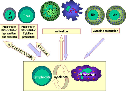
Cytokine Network
Although the focus has been on the production and action of
cytokines on cells of the immune system, it is important to understand
that many of them have effects on other cells and organ systems.
A schematic diagram showing some of the interactions in the
cytokine network is presented in Figure 9.
|
|
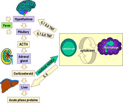
 Figure 9a
Cytokine network. Communication between lymphocytes and macrophages
and other components of the immune system
 Figure 9b Cytokine network. Communication between lymphocytes and macrophages
and the hypothalamus, adrenals and the liver
Figure 9b Cytokine network. Communication between lymphocytes and macrophages
and the hypothalamus, adrenals and the liver
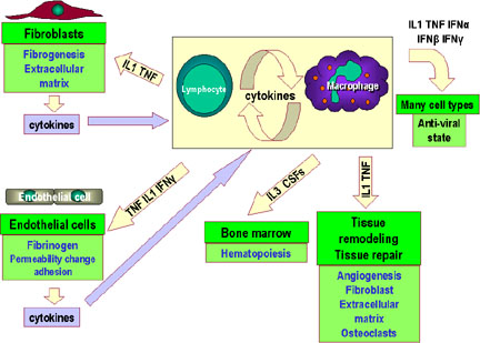
 Figure 9c
Cytokine network. Communication between lymphocytes and macrophages
and other cells and tissues
Figure 9c
Cytokine network. Communication between lymphocytes and macrophages
and other cells and tissues |
|
Table
2 - FEATURES OF CYTOKINES
|
|
Cytokine |
Cell
Source |
Cell
Target |
Primary
Effects |
|
IL-1 |
Monocytes
Macrophages
Fibroblasts
Epithelial cells
Endothelial cells
Astrocytes |
T cells; B cells
Endothelial cells
Hypothalamus
Liver
|
Costimulatory molecule
Activation
(inflammation)
Fever
Acute phase reactants
|
|
IL-2 |
T cells; NK cells
|
T cells
B cells
Monocytes
|
Growth
Growth
Activation
|
|
IL-3 |
T cells |
Bone marrow progenitors |
Growth and
differentiation |
|
IL-4 |
T cells |
Naive T cells
T cells
B cells
|
Differentiation into a
TH 2 cell
Growth
Activation and growth;
Isotype switching to IgE
|
|
IL-5 |
T cells |
B cells
Eosinophils
|
Growth and activation |
|
IL-6 |
T cells; Macrophages;
Fibroblasts |
T cells; B cells
Mature B cells
Liver
|
Costimulatory molecule
Growth (in humans)
Acute phase reactants
|
|
IL-8 family
|
Macrophages; Epithelial
cells; Platelets |
Neutrophils |
Activation and
chemotaxis |
|
IL-10 |
T cells (TH2) |
Macrophages
T cells
|
Inhibits APC activity
Inhibits cytokine
production
|
|
IL-12 |
Macrophages; NK cells |
Naive T cells |
Differentiation into a
TH 1 cell |
|
IFN-gamma |
T cells; NK cells |
Monocytes
Endothelial cells
Many tissue cells - especially macrophages
|
Activation
Activation
Increased class I and
II MHC
|
|
TGF-beta |
T cells; Macrophages |
T cells
Macrophages
|
Inhibits activation and
growth
Inhibits activation
|
|
GM-CSF |
T cells; Macrophages;
Endothelial cells, Fibroblasts |
Bone marrow progenitors |
Growth and
differentiation |
|
TNF-alpha |
Macrophages; T cells |
Similar to IL-1 |
Similar to IL-1 |
|
IL = interleukin GM-CSF
= granulocyte-macrophage colony stimulating factor
IFN = interferon TNF =
tumor necrosis factor
TGF = transforming
growth factor
|
|
|

|
 Return to the Department of Microbiology and Immunology Site Guide
Return to the Immunology Section of Microbiology and Immunology On-line
This page copyright
2004, The
Board of Trustees of the University of South Carolina
This page last changed on
Friday, July 16, 2004
Page maintained by Richard Hunt
URL: http://www.med.sc.edu:85/bowers/cell-mediated.htm
Please report any problems to rhunt@med.sc.edu
|
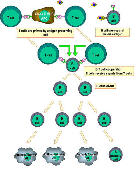 Figure 2
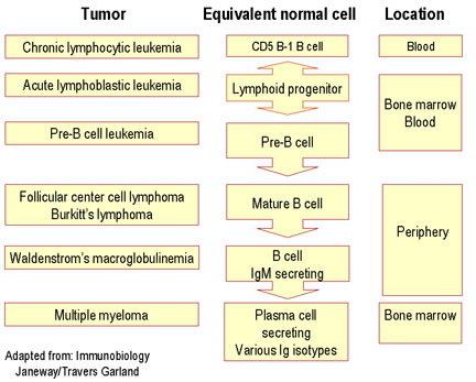 Figure 4
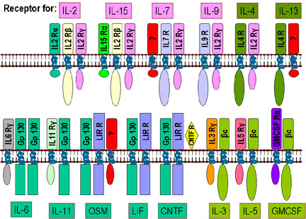 Figure 5 Receptors for various cytokines showing common subunits
 Figure 6
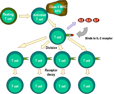 Figure 7
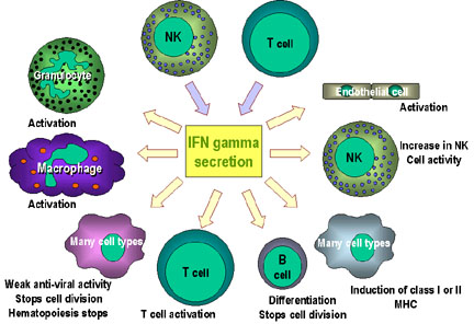 Figure 8