xx x

 INFECTIOUS DISEASE BACTERIOLOGY IMMUNOLOGY MYCOLOGY PARASITOLOGY VIROLOGY

TURKISH

   
BACTERIOLOGY - CHAPTER TEN 

General aspects of bacterial pathogenesis 

Dr Alvin Fox
Emeritus Professor
University of South Carolina School of Medicine


SPANISH

ALBANIAN
 
Let us know what you think
FEEDBACK
SEARCH
  

Logo image © Jeffrey Nelson, Rush University, Chicago, Illinois  and The MicrobeLibrary


Key Words
Pathogen/Epidemic
Normal flora
Infection
Infectious disease
Compromised host
Opportunistic infection
Nosocomial
Transmission
Koch’s postulates
Adhesion
Penetration
Invasiveness/spread
Extra/intracellular parasite
Capsule
Exotoxin
Endotoxin
Immunopathology
Autoimmunity


Pathogenesis is a multi-factorial process which depends on the immune status of the host, the nature of the species or strain (virulence factors) and the number of organisms in the initial exposure.

A limited number of bacterial species are responsible for the majority of infectious diseases in healthy individuals. Due to the success of vaccination, antibiotics, and effective public health measures, until recently, epidemics were felt to be a thing of the past. Due to the development of antibiotic resistant organisms, this situation is changing rapidly.

All humans are infected with bacteria (the normal flora) living on their external surfaces (including the skin, gut and lungs). We are constantly also exposed to bacteria (including air, water, soil and food). Normally due to our host defenses most of these bacteria are harmless. In compromised patients, whose defenses are weakened, these bacteria often cause opportunistic infectious diseases when entering the bloodstream (after surgery, catheterization or other treatment modalities). When initiated in the hospital, these infectious diseases are referred to as nosocomial. Some common bacteria found in the normal flora include Staphylococcus aureus, S. epidermidis and Propionibacterium acnes (found on the skin) and Bacteroides and Enterobacteriaceae found in the intestine (the latter in much smaller numbers).


Koch's postulates (modified)

1. The organism must always be found in humans with the infectious disease but not found in healthy ones.

2. The organism must be isolated from humans with the infectious disease and grown in pure culture.

3. The organism isolated in pure culture must initiate disease when re-inoculated into susceptible animals.

4. The organism should be re-isolated from the experimentally infected animals.

Postulates 3. and 4. are extremely important in definite proof of the role of agent in human disease. However, this depends on the ability to develop animal models that resemble the human disease. In many cases such models do not exist.

Transmission

Specific bacterial species (or strains within a species) initiate infection after being transmitted by different routes to specific sites in the human body. For example, bacteria are transmitted in airborne droplets to the respiratory tract, by ingestion of food or water or by sexual contact.

Adhesion

Bacterial infections are usually initiated by adherence of the microbe to a specific epithelial surface of the host. Otherwise the organism is removed e.g. by peristalsis and defecation (from the gut) ciliary action, coughing and sneezing (from the respiratory tract) or urination (from the urogenital tract). Adhesion is not non-specific "stickiness". Specific interactions between external constituents on the bacterial cell (adhesins) and on the host cell (receptors) occur i.e. an adhesin-receptor interaction.

S. pyogenes has surface fimbriae which contain two major components the M protein and lipoteichoic acid. The protein fibronectin binds to epithelial cells and fatty acid moieties of lipoteichoic acid in turn interact with fibronectin.

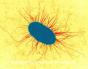
   E. coli with fimbriae (TEM x17,250)  © Dennis Kunkel Microscopy, Inc.  Used with permission Strains of E. coli with different surface characteristics cause distinct diseases. Among the most thoroughly studied pili are those of uropathogenic E. coli. Certain adhesins present on the tips of fimbriae of E. coli facilitate binding to epithelial cells. Type 1 fimbriae bind to mannose containing receptors. Whilst P fimbriae allow binding to galactose containing glycolipids (e.g. cerebrosides) and glycoproteins present on epithelial cells. They are referred to as "P" fimbriae since they were originally shown to bind to P blood group antigens on human erythrocytes.


Penetration and spread

Some bacterial pathogens reside on epithelial surfaces e.g. Vibrio cholerae. Other species are able to penetrate these barriers but remain locally. Others pass into the bloodstream or from there onto other systemic sites. This often occurs in the intestine, urinary tract and respiratory tract, and much less commonly through the skin. For example, Shigella penetrates by activating epithelial cells of the intestine to become endocytic; the Shigella do not usually spread into the bloodstream. In other cases, bacteria (e.g. Salmonella typhi) pass through epithelial cells into the bloodstream. Thus, invasion can refer to the ability of an organism to enter a cell, although in some instances it can mean further passage into the systemic vasculature. Borrelia burgdorferi is transmitted into the bloodstream through the skin by a tick bite. Certain degradative exotoxins secreted by some bacteria (e.g. hyaluronidase or collagenase) can loosen the connective tissue matrix increasing the ease of passage of bacteria through these sites.

Survival in the host

Many bacterial pathogens are able to resist the cytotoxic action of plasma and other body fluids involving antibody and complement (classical pathway) or complement alone (alternate pathway) or lysozyme. Killing of extracellular pathogens largely occurs within phagocytes after opsonization (by antibody and/or complement) and phagocytosis. Circumvention of phagocytosis by extracellular pathogens is thus a major survival mechanism. Capsules (many pathogens), protein A (S. aureus) and M protein (S. pyogenes) function in this regard.

Protein A is a surface constituent of S. aureus as well as a secreted product and binds to the Fc portion of immunoglobulins. Bacteria, on binding antibody, activate the classical complement cascade which results in the attachment of fragments of C3. Phagocytosis occurs after binding of the opsonized bacteria to receptors for the Fc portion of IgG or C3 regions. Protein A is anti-complementary (since, on binding to IgG, the complement cascade is activated, depleting complement levels). Thus in the presence of protein A, interaction of bacteria (via bound complement) with C3 receptors will be inhibited. Free protein A binds to the Fc portion of IgG, thus phagocytosis via Fc receptors may not occur because of steric hindrance.

Peptidoglycan, like lipopolysaccharide, can activate the alternate complement cascade. In S. pyogenes peptidoglycan is sufficiently exposed that it is able to bind complement. The M protein of group A streptococci is the anti-phagocytic component of the fimbriae. M protein binds fibrinogen from plasma which blocks complement binding to the underlying peptidoglycan layer. Thus streptococci in non-immune serum are not phagocytosed.

Intracellular pathogens (both obligate and facultative) must be able to avoid being killed within phagolysozomes. This can occur from by-passing or lysing these vesicles and then residing free in the cytoplasm. Alternatively, they can survive in phagosomes (fusion of phagosomes with lysosomes may be inhibited or the organism may be resistant to degradative enzymes if fusion with lysosomes occurs).

Tissue injury

Bacteria cause tissue injury primarily by several distinct mechanisms involving:

  • Exotoxins
  • Endotoxins and non-specific immunity
  • Specific humoral and cell mediated immunity


Exotoxins

Many bacteria produce proteins (exotoxins) that modify, by enzymatic action, or otherwise destroy certain cellular structures. Effects of exotoxins are usually seen acutely, since they are sufficiently potent that serious effects (e.g. death) often result. Examples of this are botulism, anthrax, cholera and diphtheria. If the host survives the acute infection, neutralizing antibodies (anti-toxins) are often elicited that neutralize the affect of the exotoxin. Classes of exotoxins include:

Toxins that act on the extracellular matrix of connective tissue

e.g. Clostridium perfringens collagenase, Staphylococcus aureus hyaluronidase.

Toxins that have a cell binding "B" component and an active "A" enzymatic component (A-B type toxins)

These include:
a) Those with ADP-ribosylating activity e.g. cholera toxin, E. coli heat labile toxin, Pseudomonas aeruginosa and diphtheria toxins.
b) Those with a lytic activity on 28S rRNA e.g. shiga and shiga-like (vero) toxins.
c) Those with a partially characterized site of action e.g. botulinum toxin, tetanus toxin and anthrax lethal toxin.

Membrane Damaging Toxins e.g. Staphylococcus aureus delta toxin

Toxins which act extracellularly. These include proteases, collagenases and hyaluronidases. For example, Clostridium perfringens produces a potent collagenase, whilst Staphylococcus aureus produces a hyaluronidase. Damage to the connective tissue matrix (by hyaluronidase and collagenase) can "loosen up" the tissue fibers allowing the organism to spread through the tissues more readily. Also included in this group is the exfoliatin of Staphylococcus aureus which causes separation of the layers within the epidermis and is the causative agent of scalded skin syndrome in the newborn.

A - B Toxins. Such toxins consist of two components. One binds to cell surfaces and the other passes into the cell membrane or cytoplasm where it acts. The classical toxins demonstrated to act in this fashion are those of cholera and diphtheria.

(i) ADP-ribosylating exotoxins

Diphtheria toxin (produced by Corynebacterium diphtheriae) is coded by the phage tox gene. The toxin is synthesized as one polypeptide chain and readily nicked into two chains held together by a disulfide bond. B binds to cells and A has the enzymatic activity. A is endocytosed and from the endosome passes into the cytosol. Diphtheria toxin ADP-ribosylates elongation factor (EF2) in ribosomes, thus inhibiting protein synthesis. Pseudomonas exotoxin A has an similar mode of action to diphtheria toxin.

Cholera toxin has several subunits which form a ring with one A subunit inserted in the center. B binds to gangliosides on the cell surface and appear to provide a channel through which A penetrates. A1 is formed by proteolytic cleavage and after internalization ADP-ribosylates a cell membrane regulator complex (using NADH as a substrate), in turn causing activation of adenylate cyclase. Activation of adenylate cyclase causes an increase in cyclic AMP production with resulting decrease in sodium chloride uptake from the lumen of the gut and active ion and water secretion with a watery diarrhea resulting. E. coli labile toxin has a similar mode of action.

(ii) Toxins that act on 28S rRNA

Shiga toxins (chromosomally encoded) are involved in the pathogenesis of shigellosis, whilst shiga-like toxins (phage encoded) are primarily produced by enterohemorraghic E. coli. They share a common mode of action. A fragment of the A subunit passes to the ribosome where it has N-glycosidase activity on a single adenosine residue; i.e. the bond between the base and ribose is lysed. Diarrhea results not from active ion/water secretion, but poor water absorption due to death of epithelial cells from inhibition of protein synthesis.

(iii) Partially characterized site of action

Botulinum neurotoxins, tetanospasmin and the lethal toxin of B. anthracis appear to be A-B type exotoxins. Botulinum toxin acts by causing inhibition of release of acetylcholine at the neuromuscular junction. Tetanus toxin is taken up at neuromuscular junctions and transported in axons to synapses. It then acts by inactivating inhibitory neurons. The exotoxins of tetanus and botulism appear to have B components, but the mode of action of their A subunits are not known. The B component of lethal toxin of B. anthracis is the protective antigen; interestingly, this also serves as the B subunit for edema toxin.

Membrane Damaging Toxins: These toxins enzymatically digest the phospholipid (or protein) components of membranes or behave as detergents. In each case holes are punched in the cell membrane and the cytoplasmic contents can leach out. The phospholipase ("toxin") of C. perfringens is an example of a membrane damaging toxin. It destroys blood vessels stopping the influx of inflammatory cells. This also helps create an anaerobic environment which is important in the growth of this strict anaerobe. The delta toxin of S. aureus is an extremely hydrophobic protein that inserts into cell membranes and is believed to have a detergent-like action.


Endotoxins

Despite the advances of the antibiotic era, around 200,000 patients will develop Gram negative sepsis each year of whom around 25-40% will ultimately die of septic shock. Septic shock involves hypotension (due to tissue pooling of fluids), disseminated intravascular coagulation and fever and is often fatal from massive system failure. This includes lack of effective oxygenation of sensitive tissues such as the brain. There is no effective therapy to reverse the toxic activity of lipid A or peptidoglycan in patients.

Endotoxins are toxic components of the bacterial cell envelope. The classical and most potent endotoxin is lipopolysaccharide. However, peptidoglycan displays many endotoxin-like properties. Certain peptidoglycans are poorly biodegradable and can cause chronic as well as acute tissue injury. Endotoxins are "non-specific" inciters of inflammation. For example, cells of the immune system and elsewhere are stimulated to release cytokines (including interleukin 1 and tumor necrosis factor). Endotoxins also activate the alternate complement pathway. The production of these cytokines results in attraction of polymorphonuclear cells into affected tissues. PG and LPS and certain other cell wall components (e.g. pneumococcal teichoic acid) are also activators of the alternate complement cascade. Thus many bacteria will bind complement encouraging their uptake and killing by phagocytes in the absence of antibody. Certain complement by-products are also chemoattractants for neutrophils. Endotoxins are also potent B cell mitogens, polyclonal B cell activators and adjuvants (for both antibodies and cell mediated immunity); this plays a role in the development of a suitable chronic immune response in handling the microbes if they are not eliminated acutely.

In a "primary" infection during the acute phase "non-antigen specific" immunity will be of utmost importance in eradicating the infection. If the organism persists (or in a reinfection at a later date), specific immunity will be of greater significance in slowing growth of the organisms or in eliminating infection. This is important in chronic infections such as tuberculosis, leprosy, Lyme disease and syphilis.


Immunopathology

The infected tissue often serves as an innocent bystander and immunopathology results. This can occur in acute and chronic infections. Over stimulation of cytokine production and complement activation by endotoxins can cause tissue injury in the absence of an immune response. Continuously generated antigens released from persisting viable microbes will subsequently elicit humoral antibodies and cell mediated immunity resulting in chronic immunopathology. Certain poorly degradable antigens (e.g pneumococcal polysaccharide and group A streptococcal cell walls) can maintain immunopathology even in the absence of persistence of live agents. Other bacterial antigens cross-react with host tissue antigens causing the development of autoimmunity (e.g. the M protein of S. pyogenes cross-reacts with mammalian myosin). Thus immunopathology can persist even after the infection and microbial antigens are eliminated.

The immune system in resistance to infection - examples

1. Extracellular parasites. Antibodies cause lysis of the organism and/or their opsonization by phagocytes at which point they are rapidly killed.

2. Intracellular parasites are primarily killed by cell mediated immunity.

3. Exotoxins can be neutralized by antitoxins. These can be elicited using toxoid vaccines (toxoids are antigenic but not toxic). This occurs, for example, in vaccination against diphtheria.

4. Certain organisms produce IgA proteases (including H. influenzae, S. pneumoniae, N. gonorrhoeae and N. meningitidis) this helps survival on external surfaces.


Some Organisms of Medical Interest

Gram negative aerobic cocci
Neisseria

Gram positive cocci (facultative anaerobes)
Streptococcus

Staphylococcus

Spirochetes
Treponema
Borrelia
Leptospira

Gram positive anaerobic cocci
Peptococcus
Peptostreptoccus

Spiral, Gram negative
Campylobacter
Helicobacter

Endospore forming Gram positive rods
Bacillus
(aerobic)
Clostridium
(anaerobic)

Gram negative aerobic rods
Pseudomonas
Bordetella
Francisella

Gram positive asporogenous aerobic rods
Listeria
Erysipelothrix

Gram negative facultative rods
a)
Enterobacteriaceae
Escherichia
Salmonella
Shigella
Yersinia
Enterobacter
Proteus
Serratia
Edwardsiella

b) Others
Vibrio
Hemophilus
Pasteurella

c) Legionellaceae
Legionella
Tatlockia


Actinomycetes and related organisms
Corynebacterium
Mycobacterium
Nocardia
Actinomyces

Corynebacterium-like in appearance
Propionibacterium

Fastidious Gram negative bacteria
Brucella
 Rochalimeae/Bartonella
Chlamydia
Rickettsia
Mycoplasma

Gram negative anaerobic rods
Bacteroides


SOME MAJOR EXOTOXINS

  Organism Disease Toxin Further Information
Bacillus anthracis Anthrax

Edema toxin

Edema factor/protective antigen complex
    Lethal toxin Lethal factor/protective antigen complex
Clostridium botulinum Botulism Botulism .toxin Blocks release of acetylcholine
Clostridium difficile

Pseudo  membranous colitis

Enterotoxin
Clostridium perfringens Gas gangrene

Alpha toxin  Hyaluronidase

Phospholipase,  (lecithinase)
  Food poisoning Enterotoxin  
Clostridium tetani Tetanus

Tetanospasmin

Blocks action of inhibitory neurones
Corynebacterium diphtheriae Diphtheria Diphtheria toxin  Inhibits elongation factor- 2 (EF2) by ADP ribosylation
Escherichia coli Diarrhea (ETEC)

Heat labile toxin 

Activates adenyl cyclase
    Heat stable toxins  Activates adenyl cyclase
  Hemorrhagic colitis

Vero toxin

 
Pseudomonas aeruginosa Diseases of compromised host Exotoxin A  Inhibits EF2
Staphylococcus aureus Opportunistic infections Alpha-gamma toxins, leucocidin
Toxic shock  Toxic shock toxin
Food poisoning Enterotoxin
Scalded skin syndrome Exfoliatin
Streptococcus pyogenes Scarlet fever
Toxic shock
Erythrogenic/pyrogenic toxin   
  Shigella dysenteriae  

Bacillary dysentery

Shiga toxin

Inhibits protein synthesis by lysing 28S rRNA
Vibrio cholerae Cholera Choleragen Activates adenyl cyclase by ADP-ribosylation


Return to the Bacteriology Section of the Microbiology and Immunology On-line Textbook


This page last changed on Monday, February 29, 2016
Page maintained by
Richard Hunt