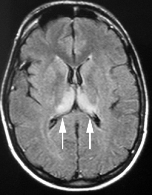
The MRI is generally the most useful supportive diagnostic test in variant CJD. It is a relatively non invasive investigation that is generally readily available and, importantly, is undertaken in cases of suspect variant CJD in order to exclude other possible illnesses. However, there is a characteristic abnormality seen in the posterior thalamic region (the so called “pulvinar sign”) which is highly sensitive and specific for variant CJD. The pulvinar sign has been found in 90+% of pathologically proven vCJD cases. Present indications are that FLAIR sequences are most likely to show the abnormality. The stage of illness at which the scan becomes positive is uncertain. However, individual patients generally have MR scans at a time when clinicians feel they are appropriate and, as indicated above, partly to exclude other illnesses. It is at this clinically indicated point that positive scans have been found to be positive. In a few patients, the pulvinar sign has been present on MRI within 3 months of symptom onset.
The finding of a positive MRI scan allows a “possible variant CJD” case to be elevated to the category of “probable variant CJD”.
Figure: Transverse FLAIR MRI showing bilateral and symmetrical high signal in the pulvinar nuclei of the thalamus - the 'pulvinar sign' of variant CJD.