|
|
 |
|||||
|
Dr Richard Hunt |
BACTERIOLOGY | IMMUNOLOGY | MYCOLOGY | PARASITOLOGY | VIROLOGY | |
|
|
PICORNAVIRUSES - PART TWO RHINOVIRUSES
|

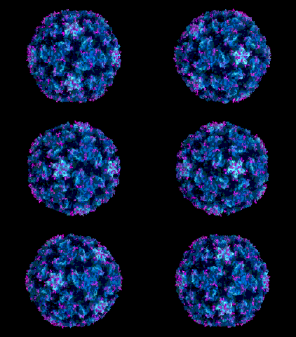
Image © Jean-Yves
Sgro. Institute for Molecular |
||||
|
WEB RESOURCES |
Rhinoviruses (Rhinos - nose (Greek))
are one of the families of viruses that can cause the common cold although
many other viruses can infect the respiratory tract and cause cold-like
symptoms. It is estimated that about one third of "colds" are caused
by rhinovirus infections. There are more than 100 serotypes explaining why
vaccines against rhinoviruses have proved difficult to develop. Rhinoviruses
have a diameter of about 30nm and are
positive strand RNA viruses with a naked nucleocapsid (figure 1). They are sensitive to low
pH and, as might be expected from their symptoms, are spread by aerosols and
infect the upper respiratory tract. They can also be spread by fomites
such as hands and other forms of direct contact. Rhinoviruses are quite stable,
lasting for hours on fomites, but are sensitive to temperature. Thus, they do not spread to the lower respiratory tract since they replicate best at a
few degrees below normal body temperature. Although the most common route
of infection is the nose, virus can also enter via the mouth and the eyes. There
is usually no gastrointestinal involvement because of the acid lability
of the virus. The virus is therefore not spread from the intestinal
tract.
RHINOVIRUS DISEASE
The symptoms of a rhinovirus infection are well known: discharging or blocked nasal passages often accompanied by sneezes, and perhaps a sore throat. This typical "runny nose" (rhinorhea) may be accompanied by a general malaise, cough, sore throat etc. The characteristic symptoms occur from one to four days after infection at which time extremely high titers of the rhinovirus are found in the nasal secretions (there can be as many as 1000 infectious virus particles per ml). It appears that one rhinovirus infectious virion particle is capable of initiating disease. The virus replicates itself primarily in epithelial cells of the nasal mucosa but there is little damage to the mucosa although infected cells may be sloughed off. There may be edema of connective tissue. The symptoms experienced depend on the number of virus particles replicated. Infected cells produce a variety of molecules that such as histamine that result in increased nasal secretions. It is the production of such molecules rather than direct cellular destruction to accounts for the symptoms experienced by the patient. These molecules cause changes in vascular permeability The primary infection results in IgA in nasal secretions and IgG in the bloodstream. Since these viruses do not enter the circulation, the mucosal IgA response is the most important. This leads to immunity and resolution of the disease although the levels of nasal IgA are rapidly reduced. Immunity against a particular serotype may last 1 to 2 years but as noted above there are many serotypes against which protection is not gained. As with infections by other viral infections, interferon production is the primary means of defense, preceding the antibody response. Interferon production may lead to the symptoms experienced by the patient (see Virus-Host Interactions). Many infected persons (about 50%) do not show symptoms of a rhinovirus infection but are nevertheless capable of passing on the infection. Although the lower respiratory tract is usually not affected, bronchopneumonia can occur in rhinovirus infections, particularly in children. |
|||||
 Figure 1 Human rhino virus © Dr J-Y Sgro, University of
Wisconsin. Used with permission
 |
||||||
|
EPIDEMIOLOGY Rhinovirus infections usually occur at times of increased human contact, that is in the colder months of the year. Many different serotypes circulate simultaneously. Frequently children become infected and then pass the virus to adults after an incubation time of about two or three days. Often as many as one half of the contacts get a cold in this way. Antigenic variation occurs. Many infections by other viruses cause symptoms that are similar to those of rhinoviruses. These include parainfluenzaviruses, coronaviruses and enteroviruses RECEPTORS Most rhinoviruses bind to a member of the immunoglobulin super-family of proteins, ICAM-1 which is found on the surfaces of epithelial and other cells. This molecule mediates cell-cell adhesion in a variety of epithelial cells. The expression of ICAM-1 is enhanced under inflammatory conditions such as occur in a rhinovirus infection which may lead to viral spread because of more available receptor molecules (positive feedback loop). Because of the specificity to ICAM-1, only humans are infected by human rhinoviruses.
|
||||||
|
|
CULTURE If it is necessary to identify the virus that gives rise to the typical "cold" symptoms, virus can be grown on cultured cells from nasal specimens. Usually, human fibroblasts are used and the laboratory looks for a typical cytopathic effect of refractile cells (figure 2). The cells are grown at around 33 degrees. DIAGNOSIS Many types of viruses give "cold"-like symptoms and it is usually unnecessary to carry out further identification. Usually it is enough to note the minor symptoms and the seasonal infections. There are specific antibody tests available but these are not generally used. TREATMENT There is usually no need to treat the infection although treatment of the symptoms may be used. This often consists of rehydration and keeping the airways unblocked. Physicians often prescribe aspirin to relive fever symptoms but this may exacerbate viral proliferation if body temperature is reduced since, as noted above, the virus is particularly temperature-sensitive. Interferon nasal sprays have little effect. Pleconaril is broadly active against rhinoviruses (see Anti-Viral Chemotherapy). The best way to avoid spreading the virus is interrupt the infection chain by hand washing.
|
|||||
|
This
page copyright 2005, The Board of Trustees of the University of South
Carolina |
||||||