|
x |
x |
|
 |
 |
|
INFECTIOUS
DISEASE |
BACTERIOLOGY |
IMMUNOLOGY |
MYCOLOGY |
PARASITOLOGY |
VIROLOGY |
|
|
BACTERIOLOGY - CHAPTER SEVEN
VIROLOGY - CHAPTER TWENTY
FOUR
BACTERIOPHAGE
Gene Mayer, PhD
Professor Emeritus
Department of Pathology, Microbiology and Immunology
University of South Carolina School of Medicine
Columbia
|
|
SPANISH |
|
FRENCH |
|
ALBANIAN |
|
PORTUGUESE |
Let us know what you think
FEEDBACK |
|
SEARCH |
|
|
|

Bacteriology
Virology

Bacteriology
Virology
|
|
Logo image © Jeffrey
Nelson, Rush University, Chicago, Illinois and
The MicrobeLibrary |
|
TEACHING OBJECTIVES
To describe the general composition and structure of bacteriophage
To discuss the infectious process and the lytic
multiplication cycle
To explain the lysogenic cycle and its regulation
 © CellsAlive
- James A. Sullivan
© CellsAlive
- James A. Sullivan |
INTRODUCTION
Bacteriophage (phage) are obligate
intracellular parasites that multiply inside bacteria by making use of some
or all of the host biosynthetic machinery (i.e., viruses that infect
bacteria.).
There are many similarities between bacteriophages and
animal cell viruses. Thus, bacteriophage can be viewed as model systems for
animal cell viruses. In addition a knowledge of the life cycle of
bacteriophage is necessary to understand one of the mechanisms by which
bacterial genes can be transferred from one bacterium to another.
At one time it was thought that the use of bacteriophage
might be an effective way to treat bacterial infections, but it soon became
apparent that phage are quickly removed from the body and thus, were of
little clinical value. However, bacteriophage are used in the diagnostic
laboratory for the identification of pathogenic bacteria (phage typing).
Although phage typing is not used in the routine clinical laboratory, it is
used in reference laboratories for epidemiological purposes. Recently, new
interest has developed in the possible use of bacteriophage for treatment of
bacterial infections and in prophylaxis. Whether bacteriophage will be used
in clinical medicine remains to be determined.
|
|
KEY WORDS
Bacteriophage
Phage
typing
Capsid
Tail
Contractile
sheath
Base plate
Tail
fibers
Virulent
phage
Eclipse
Early and late m-RNA
Plaque
Pfu
Lysogeny
Temperate
phage
Prophage
Lysogen
Cohesive
ends
Site-specific
recombination
Repression
Induction
Lysogenic conversion
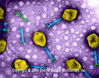
T4 Bacteriophage (TEM x390,000) ©
Dennis Kunkel Microscopy, Inc.
Used with permission

T4 bacteriophage Negative stain electron micrograph
©
ICTV.
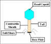 Figure 1 Structure of T4 bacteriophage
 |
COMPOSITION AND STRUCTURE OF BACTERIOPHAGE
Composition
Although different bacteriophages
may contain different materials they all contain nucleic acid
and protein.
Depending upon the phage, the nucleic acid can be either DNA
or RNA but not both and it can exist in various forms. The nucleic acids of
phages often contain unusual or modified bases. These modified bases protect
phage nucleic acid from nucleases that break down host nucleic acids during
phage infection. The size of the nucleic acid varies depending upon the
phage. The simplest phages only have enough nucleic acid to code for 3-5
average size gene products while the more complex phages may code for over
100 gene products.
The number of different kinds of protein and the amount of
each kind of protein in the phage particle will vary depending upon the
phage. The simplest phage have many copies of only one or two different
proteins while more complex phages may have many different kinds. The
proteins function in infection and to protect the nucleic acid from
nucleases in the environment .
Structure
Bacteriophage come in many different
sizes and shapes. The basic structural features of bacteriophages are
illustrated in Figure 1, which depicts the phage called T4.
Size
T4 is among the largest phages; it is
approximately 200 nm long and 80-100 nm wide. Other phages are smaller.
Most phages range in size from 24-200 nm in length.
Head or Capsid
All phages contain a head
structure which can vary in size and shape. Some are icosahedral (20
sides) others are filamentous. The head or capsid is composed of many
copies of one or more different proteins. Inside the head is found the
nucleic acid. The head acts as the protective covering for the nucleic
acid.
Tail
Many but not all phages have tails
attached to the phage head. The tail is a hollow tube through which the
nucleic acid passes during infection. The size of the tail can vary and
some phages do not even have a tail structure. In the more complex
phages like T4 the tail is surrounded by a contractile sheath which
contracts during infection of the bacterium. At the end of the tail the
more complex phages like T4 have a base plate and one or more tail
fibers attached to it. The base plate and tail fibers are involved in
the binding of the phage to the bacterial cell. Not all phages have base
plates and tail fibers. In these instances other structures are involved
in binding of the phage particle to the bacterium.
INFECTION OF HOST CELLS
Adsorption
The first step in the infection
process is the adsorption of the phage to the bacterial cell. This step is
mediated by the tail fibers or by some analogous structure on those phages
that lack tail fibers and it is reversible. The tail fibers attach to
specific receptors on the bacterial cell and the host specificity of the
phage (i.e. the bacteria that it is able to infect) is usually
determined by the type of tail fibers that a phage has. The nature of the
bacterial receptor varies for different bacteria. Examples include proteins
on the outer surface of the bacterium, LPS, pili, and lipoprotein. These
receptors are on the bacteria for other purposes and phage have evolved to
use these receptors for infection.
Irreversible attachment
The attachment of the
phage to the bacterium via the tail fibers is a weak one and is reversible.
Irreversible binding of phage to a bacterium is mediated by one or more of
the components of the base plate. Phages lacking base plates have other ways
of becoming tightly bound to the bacterial cell.
|
|
MOVIE
Bacteriophage
Requires Quicktime
© Mondo Media
San Francisco, California 94107 USA
and The MicrobeLibrary |
 Figure 2 Contraction of the tail sheath of T4
 |
Sheath Contraction
The irreversible binding of the
phage to the bacterium results in the contraction of the sheath (for those
phages which have a sheath) and the hollow tail fiber is pushed through the
bacterial envelope (Figure 2). Phages that don't have contractile sheaths
use other mechanisms to get the phage particle through the bacterial
envelope. Some phages have enzymes that digest various components of the
bacterial envelope.
Nucleic Acid Injection
When the phage has gotten
through the bacterial envelope the nucleic acid from the head passes through
the hollow tail and enters the bacterial cell. Usually, the only phage
component that actually enters the cell is the nucleic acid. The remainder
of the phage remains on the outside of the bacterium. There are some
exceptions to this rule. This is different from animal cell viruses in which
most of the virus particle usually gets into the cell. This difference is
probably due to the inability of bacteria to engulf materials.
|
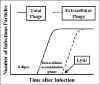 Figure 3 Life cycle of a lytic phage
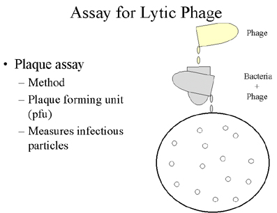
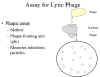 Figure 4 Assay for lytic phage
 |
PHAGE MULTIPLICATION CYCLE
Lytic or Virulent Phages
Definition
Lytic or virulent phages are
phages which can only multiply on bacteria and kill the cell by lysis at
the end of the life cycle.
Life cycle
The life cycle of a lytic phage
is illustrated in Figure 3 .
Eclipse period
During the eclipse
phase, no infectious phage particles can be found either inside or
outside the bacterial cell. The phage nucleic acid takes over the host
biosynthetic machinery and phage specified m-RNA's and proteins are
made. There is an orderly expression of phage directed macromolecular
synthesis, just as one sees in animal virus infections. Early m-RNA's
code for early proteins which are needed for phage DNA synthesis and
for shutting off host DNA, RNA and protein biosynthesis. In some cases
the early proteins actually degrade the host chromosome. After phage
DNA is made late m-RNA's and late proteins are made. The late proteins
are the structural proteins that comprise the phage as well as the
proteins needed for lysis of the bacterial cell.
Intracellular Accumulation Phase
In
this phase the nucleic acid and structural proteins that have been
made are assembled and infectious phage particles accumulate within
the cell.
Lysis and Release Phase
After a
while the bacteria begin to lyse due to the accumulation of the phage
lysis protein and intracellular phage are released into the medium.
The number of particles released per infected bacteria may be as high
as 1000.
Assay for Lytic Phage
Plaque assay
Lytic phage are
enumerated by a plaque assay. A plaque is a clear area which results
from the lysis of bacteria (Figure 4). Each plaque arises from a
single infectious phage. The infectious particle that gives
rise to a plaque is called a pfu (plaque forming unit).
|
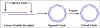 Fig. 5. Circularization of phage chromosome: cohesive ends
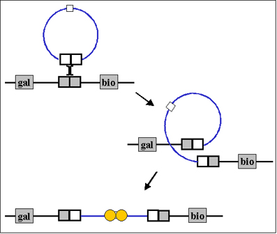
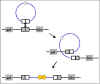 Figure 6 Site-specific recombination
 |
Lysogenic or Temperate Phage
Definition
Lysogenic or temperate phages are
those that can either multiply via the lytic cycle or enter a quiescent
state in the cell. In this quiescent state most of the phage genes are
not transcribed; the phage genome exists in a repressed state. The phage
DNA in this repressed state is called a prophage because
it is not a phage but it has the potential to produce phage. In most
cases the phage DNA actually integrates into the host chromosome and is
replicated along with the host chromosome and passed on to the daughter
cells. The cell harboring a prophage is not adversely affected by the
presence of the prophage and the lysogenic state may persist
indefinitely. The cell harboring a prophage is termed a lysogen.
Events Leading to Lysogeny
The Prototype
Phage: Lambda
Circularization of the phage chromosome
Lambda DNA is a double stranded linear molecule with small single
stranded regions at the 5' ends. These single stranded ends are
complementary (cohesive ends) so that they can base pair and
produce a circular molecule. In the cell the free ends of the circle
can be ligated to form a covalently closed circle as illustrated in
Figure 5.
Site-specific recombination
A
recombination event, catalyzed by a phage coded enzyme, occurs between
a particular site on the circularized phage DNA and a particular site
on the host chromosome. The result is the integration of the phage DNA
into the host chromosome as illustrated in Figure 6.
Repression of the phage genome
A
phage coded protein, called a repressor, is made which
binds to a particular site on the phage DNA, called the operator,
and shuts off transcription of most phage genes EXCEPT the repressor
gene. The result is a stable repressed phage genome which is
integrated into the host chromosome. Each temperate phage will only
repress its own DNA and not that from other phage, so that repression
is very specific (immunity to superinfection with the same phage).
|
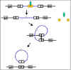 Figure 7 Termination of lysogeny
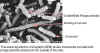 Figure 8A Scanning electron micrograph of Escherichia coli cells with phage particles (which
appear as small white dots) attached to the outside of cells.
© Scott Kachlany, Cornell University Ithaca, New York,
USA and The MicrobeLibrary
Figure 8A Scanning electron micrograph of Escherichia coli cells with phage particles (which
appear as small white dots) attached to the outside of cells.
© Scott Kachlany, Cornell University Ithaca, New York,
USA and The MicrobeLibrary
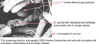 Figure 8B
SEM of E. coli cells with disrupted cell envelopes, presumably due to phage release. After the phage replicate within host cells, they must be released from the host cells. This often occurs by lysing the cell.
© Scott Kachlany, Cornell University Ithaca, New York, USA
and The MicrobeLibrary |
Events Leading to Termination of Lysogeny
Anytime a lysogenic bacterium is exposed to adverse
conditions, the lysogenic state can be terminated. This process is
called induction. Conditions which favor the termination
of the lysogenic state include: desiccation, exposure to UV or ionizing
radiation, exposure to mutagenic chemicals, etc. Adverse conditions lead
to the production of proteases (rec A protein) which destroy the
repressor protein. This in turn leads to the expression of the phage
genes, reversal of the integration process and lytic multiplication.
Lytic vs Lysogenic Cycle
The decision for lambda to enter the lytic or lysogenic
cycle when it first enters a cell is determined by the concentration of
the repressor and another phage protein called cro in the
cell. The cro protein turns off the synthesis of the repressor and thus
prevents the establishment of lysogeny. Environmental conditions that
favor the production of cro will lead to the lytic cycle while those
that favor the production of the repressor will favor lysogeny.
Significance of Lysogeny
Model for animal virus transformation
Lysogeny is a model system for virus transformation of animal cells
Lysogenic conversion
When a cell
becomes lysogenized, occasionally extra genes carried by the phage get
expressed in the cell. These genes can change the properties of the
bacterial cell. This process is called lysogenic or phage conversion.
This can be of significance clinically. e.g. Lysogenic phages
have been shown to carry genes that can modify the Salmonella O
antigen, which is one of the major antigens to which the immune
response is directed. Toxin production by Corynebacterium
diphtheriae is mediated by a gene carried by a phage. Only those
strain that have been converted by lysogeny are pathogenic.
|
|
|
 Return to the Bacteriology Section of the
Microbiology and Immunology On-line Textbook Return to the Bacteriology Section of the
Microbiology and Immunology On-line Textbook
This page last changed on
Wednesday, November 23, 2016
Page maintained by
Richard Hunt
|
 Figure 2 Contraction of the tail sheath of T4
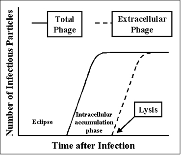 Figure 3 Life cycle of a lytic phage
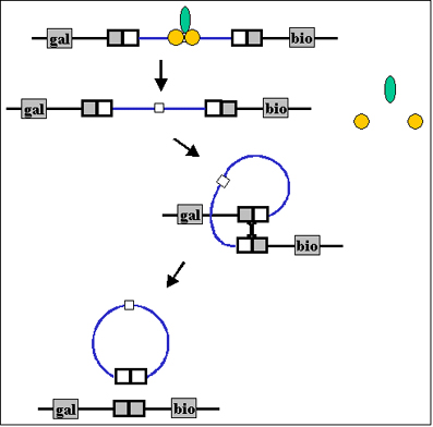 Figure 7 Termination of lysogeny