x x

INFECTIOUS DISEASE

BACTERIOLOGY IMMUNOLOGY MYCOLOGY PARASITOLOGY VIROLOGY

TURKISH


VIROLOGY - CHAPTER SEVEN    

PART NINE

HUMAN IMMUNODEFICIENCY VIRUS AND AIDS

STRUCTURE: The Genome AND PROTEINS of HIV

Dr Richard Hunt
Professor
Department of Pathology, Microbiology and Immunology
University of South Carolina School of Medicine

En Español
SHQIP - ALBANIAN
Let us know what you think
FEEDBACK
SEARCH
  
THIS CHAPTER IS IN SEVERAL PARTS
 USE THE NEXT>> BUTTON ABOVE TO GO TO THE NEXT PART
 
 TO CONTINUE TO VIROLOGY CHAPTER EIGHT USE LINK ABOVE
 

LINKS  TO OTHER HIV AND AIDS SECTIONS ARE AT THE BOTTOM OF THIS PAGE

This thin-section transmission electron micrograph (TEM) depicts the ultrastructural details of a number of ”human immunodeficiency virus” (HIV) virus particles, or virions
CDC/ Dr. A. Harrison; Dr. P. Feorino

hiv tm em.jpg (102765 bytes)    Electron micrograph of HIV  (click to enlarge) -  Cone-shaped cores are sectioned in various orientations. Viral genomic RNA is located in the electron-dense wide end of core. CDC/Dr. Edwin P. Ewing, Jr.  epe1@cdc.gov 


hivstruct.gif (32307 bytes) Diagram of the protein locations in HIV

 HIVmod2.GIF (50787 bytes) Cut-away diagram of HIV. From Dr. Milan V.Nermut of the National Institute for Biological Standards and Control. Herts, U.K. Computer graphics by A.Davies

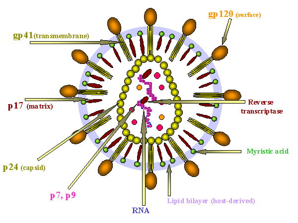
Figure  14 - HIV structure

Figure 14a
The gag gene


  Figure  24A
The genome of HIV-1


Figure 24B
The structure of gp160.
A. The linear domain structure of gp160 is shown at the top. Gp160 is cleaved into gp120 (the surface protein) and gp41 (the transmembrane fusion protein).
B. A trimer of gp120/gp41 is associated with the viral membrane.
C. Gp120 has a number of hypervariable domains (V1-V5). The red bars show disulfide bridges

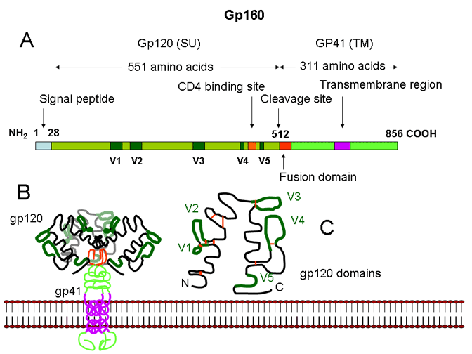
STRUCTURE OF THE VIRUS

HIV is a retrovirus with a similar structure to other retroviruses (see oncogenic viruses).

SURFACE STRUCTURES

Viral membrane

The  membrane is host-derived as a result of budding from the cell surface (figure 15). Some host proteins become incorporated into the viral membrane. This lipid envelope makes the virus susceptible to organic solvents.

Surface glycoprotein

Gp160 is encoded by the env (envelope) gene. Gp160 is cleaved after translation by host enzymes in the Golgi Body to form Gp120 (SU) and Gp41 (TM). Gp 41 is embedded in the membrane, Gp120 is not but is held to Gp41 by non-covalent interactions (figure 14). It is easily shed from the virus particle. There is a large number of sugar chains on gp120 (which may pose a problem for a vaccine). Gp120 is the protein that interacts with a receptor on the cell to be infected. Gp41 is the fusogen that is exposed after Gp120 has bound to the cell.


INTERNAL STRUCTURES

Internal structural proteins

These are all encoded by the gag (group-specific antigen) gene (figure 14a). P17 matrix (MA) protein lines the inner surface of the viral membrane to which it is attached by covalently bound myristic acid. Other proteins are associated with the nucleocapsid. The group-specific antigen is made as a polyprotein and is cleaved during or after budding of the virus by a virally-encoded protease encoded by the pol gene.

Other internal proteins

These are encoded by the pol (polymerase) gene. They are enzymes that participate in integration and replication:

  • Reverse transcriptase - copies the RNA genome into double stranded DNA. It also contains an RNase H activity that cleaves the RNA of the RNA/DNA hybrid that is formed after the first round of polymerization.

  • Integrase - integrates the double stranded DNA into the host cell chromosome

  • Protease - cleaves the pol and gag-encoded polyproteins

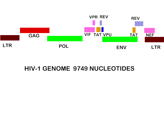
For further information on retrovirus structure and replication, see oncogenic viruses

Genome

Like other retroviruses, the genome is diploid positive sense RNA

Since HIV has a more complex life cycle that simple retroviruses such as Rous Sarcoma Virus (RSV) and it appears that HIV can control its replication in a more complex fashion, we might expect more genetic information but this is not so. The HIV genome is 9749 nucleotides - about the same size as other retroviruses, for example RSV.

The genome of HIV is more complex than RSV, however, since it has extra open reading frames that clearly code for small proteins (figure 24a). Antibodies against these small proteins are found in HIV-infected people. Some of these are protein synthesis-controlling proteins.

The HIV genome has nine open reading frames (leading to nine primary translation products) but 15 proteins are made in all as a result of cleavage of three of the primary products.

As we have already seen, the GAG gene and the GAG and POL genes together are translated into large polyproteins which are then cleaved by a virus-encoded protease that is part of the POL polyprotein. 

GAG polyprotein  is cleaved to into four proteins that are found in the mature virus:  MA (matrix), CA (capsid), NC (nucleocapsid), p6

More on MA

More on CA More on NC More on P6


POL polyprotein is cleaved to three proteins: PR (protease), RT (reverse transcriptase), IN (integrase)

More on PR More on RT  More on IN


ENV gene is translated to a polyprotein (Gp160) which is then cleaved by a host cell protease (called furin) that is found in the Golgi Body. It is not cleaved by the virus-encoded protease. Gp160 is cleaved to: SU (Gp120) and TM (Gp41). The latter retains the transmembrane part of Gp160 while Gp120 remains attached to Gp41 via non-covalent bonds.

More on SU

In a addition to the nine proteins derived from  GAG, POL and ENV, there are six other proteins made by HIV. Three of these are incorporated into the virus (Vif,  Vpr and Nef), while the others are not found in the mature virus: Tat and Rev are regulatory proteins and Vpu indirectly assists in assembly. The genes that encode these proteins are known by three letter names that are derived as follows:

TAT: Trans-Activator of Transcription

REV: Regulator of Virion protein expression

NEF: Negative Regulatory Factor

VIF: Virion Infectivity Factor

VPU: Viral Protein U

VPR: Viral Protein R

These genes encode small proteins; TAT, for example, consists of 88 amino acids. They overlap with the structural genes (especially ENV) but are in different reading frames. From the organization of the HIV genome shown in figure 24, it can be seen  that some are encoded in more than one exon (unlike the structural genes) and therefore their mRNAs can be derived by alternative splicing of mRNAs for the structural proteins. Mutants in the TAT and REV genes show that both proteins are necessary for virus production.


TAT

TAT gene product binds to a sequence in all of the genes of HIV and positively stimulates transcription. It is thus a positive regulator of protein synthesis, including its own synthesis.

More on TAT


REV

REV binds to an element only in the mRNA for structural proteins (GAG/POL/ENV) and regulates the ratio of structural to non-structural, controlling protein (TAT/REV) synthesis. When REV levels are high, structural protein synthesis rises and the synthesis of non-structural, controlling proteins falls. Thus, REV inhibits its own production and that of TAT.

The normal result is homeostasis, low or non-existent virus production and latency in the resting CD4 cell.

As we have seen, there is an inherent problem in HIV's lifestyle. It uses genomic RNA as its messenger RNA. This RNA is unspliced and the nucleus has a mechanism to prevent unspliced mRNAs from leaving the nucleus and being translated. It is the function of REV to overcome this problem.

More on REV


nef1.jpg (193062 bytes)

nef2.jpg (192449 bytes) Figure 25  NEF effects on CD4


nefcyt1.jpg (175863 bytes)

nefcyt2.jpg (218688 bytes) Figure 26   NEF induces cytokines that attract T cells to an infected macrophage

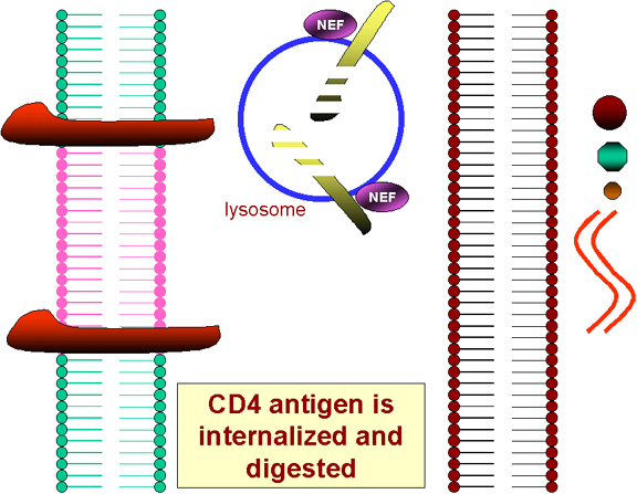
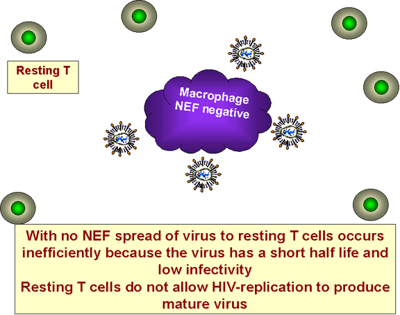
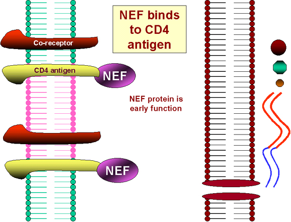
NEF

NEF protein is synthesized early in infection. Despite its small size, NEF has several functions.

  • Homeostasis leads to problems for the parasitic provirus: 

  • Super-infection by other HIV particles which bind to surface CD4 antigen of the infected cell may kill the cell

  • Probably more importantly, virus bound via CD4 antigen at the cell surface or free Gp120 bound to CD4 antigen at the cell surface may result in the cell being subject to an immune attack and the infected cell may be destroyed.

The translation of the NEF gene as a result of the first infecting virus causes the internalization of CD4 antigen from the cell surface and its destruction in lysosomes (figure 25). Thus no more HIV or gp120 can bind to the surface of an infected cell! 

  • By a different mechanism from its down regulation of CD4 antigen, NEF reduces surface expression of MHC class I molecules. This alters antigen presentation by the infected cell and is proposed to protect the infected cell from attack by cytotoxic T cells
     

  • The name, NEF, comes from negative factor. Originally, it seemed that virions that lacked NEF grew better than wild type. Now the consensus is for the opposite, that is that virus produced in the presence of NEF is a little more infectious than virus produced in its absence.
     

  • NEF is important for HIV replication in vivo but there seems to be much less effect of NEF in an in vitro cell culture situation. Why this is so has long been obscure. Recently, this question seems to have been solved. The answer is found in the macrophages which alter their secretion properties when they are infected with a NEF-expressing HIV (remember that macrophages are the cells that bring HIV into the body and the initial strains of HIV in an infected patient are macrophage-tropic).  

  • HIV-infected macrophages expressing NEF secrete MIP-1alpha and MIP-1beta. These are two chemokines that  bind to the co-receptors for HIV infection of macrophages but here these they have another function. They cause resting CD4+ T cells to migrate (undergo chemotaxis) towards the infected macrophages (figure 26). This is important in vivo since, initially, HIV-infected cells are not very numerous and uninfected T cells may not be in the vicinity of the infected cells. Moreover, HIV does not have a very long half life in the circulation before becoming non-infectious. Migration of uninfected cells towards infected cells increases the probability that the T cells will encounter infected macrophages before they leave the reticuloendothelial system. This explains why NEF seems not to be of much consequence in cell culture where the cells are already close together.
     

  • The HIV-infected macrophages expressing NEF do something else. They make a factor that has not yet been identified that activates the resting T cells that have been attracted towards them allowing the T cells to be productively infected and to shed new virus.

 These findings explain why macrophages are vital for the spread of HIV.

Note that, in vivo, HIV can infect a resting T cell but cannot replicate in that cell. Although NEF can activate the cell, it cannot be made in the resting T cell. The above observations solve this conundrum. Macrophages are infected by HIV and make NEF without any activation process. As  a result they make factors that activate resting T cells that can now support a productive infection!

More on NEF


VPU 

After activation of the T cell, the virus faces another problem: CD4 antigen and Gp120 are being made in the endoplasmic reticulum of the same cell. They are likely to bind to one another before reaching the plasma membrane and such complexes are usually targeted by the cell for degradation. To stop this unfortunate state of affairs, another of the small HIV proteins, VPU, promotes the proteolysis of the CD4 antigen of the host cell as it is made!

VPU also enhances viral particle release from the host cell. How it does this is not clear but VPU forms an ion channel in the plasma membrane of the host cell and may alter the ionic composition of the cytoplasm since it conducts small ions such as Na+ and K+. It also binds to a cellular protein (VPU-binding protein or UBP) and over-expression of this protein diminishes the enhancing effect of VPU on virus release. UBP may be a negative factor for assembly that must be displaced from one of the GAG proteins before virus can assemble at the cell surface.

From its ability to stimulate viral release and also to break down CD4 antigen (which are separate functions of different parts of the VPU molecule), it appears that VPU enhances the pathogenicity of the virus by increasing the number of HIV particles per cell.

 More on VPU


VIF 

VIF (viral infectivity factor) protein, which is essential for infection in vivo, may be very important in suppressing resistance to HIV infection by the host. VIF is needed during late stages of virus production and seems to function by suppressing innate anti-viral activities in T cells and macrophages, the major cells that are infected in humans. Without VIF, HIV is not infectious in primary human T cells. 

What is it about the T cells that render them active against HIV when VIF is absent? It is thought that VIF is needed for production of infectious virus because it inhibits an antiviral pathway in the cells that involves an enzyme called APOBEC3G (originally discovered as an apolipoprotein B messenger RNA editing enzyme). This enzyme is a cytidine deaminase that also targets single stranded DNA. It appears that VIF prevents the editing of the single strand form of the viral DNA that is the initial product of reverse transcription. Such editing is much more pronounced when the infecting virus has a VIF deletion. Thus, VIF prevents many mutations that would lead to changes in the structural proteins, enzymes and regulatory proteins that would otherwise result in loss of viral infectivity.

 More on VIF


VPR

VPR influences the pathogenesis of HIV and is essential for infection of macrophages, and to a lesser extent of other cells. It also activates HIV LTR-promoted transcription. It causes the arrest of host cell division in the G2 stage of the cell cycle and apoptosis of the infected cell. It acts as a cytoplasmic-nucleus shuttle protein (for the pre-integration complex through the nuclear pores). VPR is found in the serum of HIV-infected patients.

More on VPR


OTHER SECTIONS ON HIV

PART I HUMAN IMMUNODEFICIENCY VIRUS AND AIDS

PART II HIV AND AIDS, THE DISEASE

PART III COURSE OF THE DISEASE

PART IV PROGRESSION AND COFACTORS

PART V STATISTICS

PART VI  SUBTYPES AND CO-RECEPTORS

PART VII  COMPONENTS AND LIFE CYCLE OF HIV

PART VIII  LATENCY OF HIV

PART IX GENOME OF HIV

PART X  LOSS OF CD4 CELLS

PART XI   POPULATION POLYMORPHISM

APPENDIX I  ANTI-HIV VACCINES

APPENDIX II  DOES HIV CAUSE AIDS?

APPENDIX III  ANTI-HIV CHEMOTHERAPY


Return to the Virology Section of Microbiology and Immunology On-line

Return to the front page of Microbiology and Immunology On-line


This page last changed on Sunday, August 28, 2016
Page maintained by
Richard Hunt